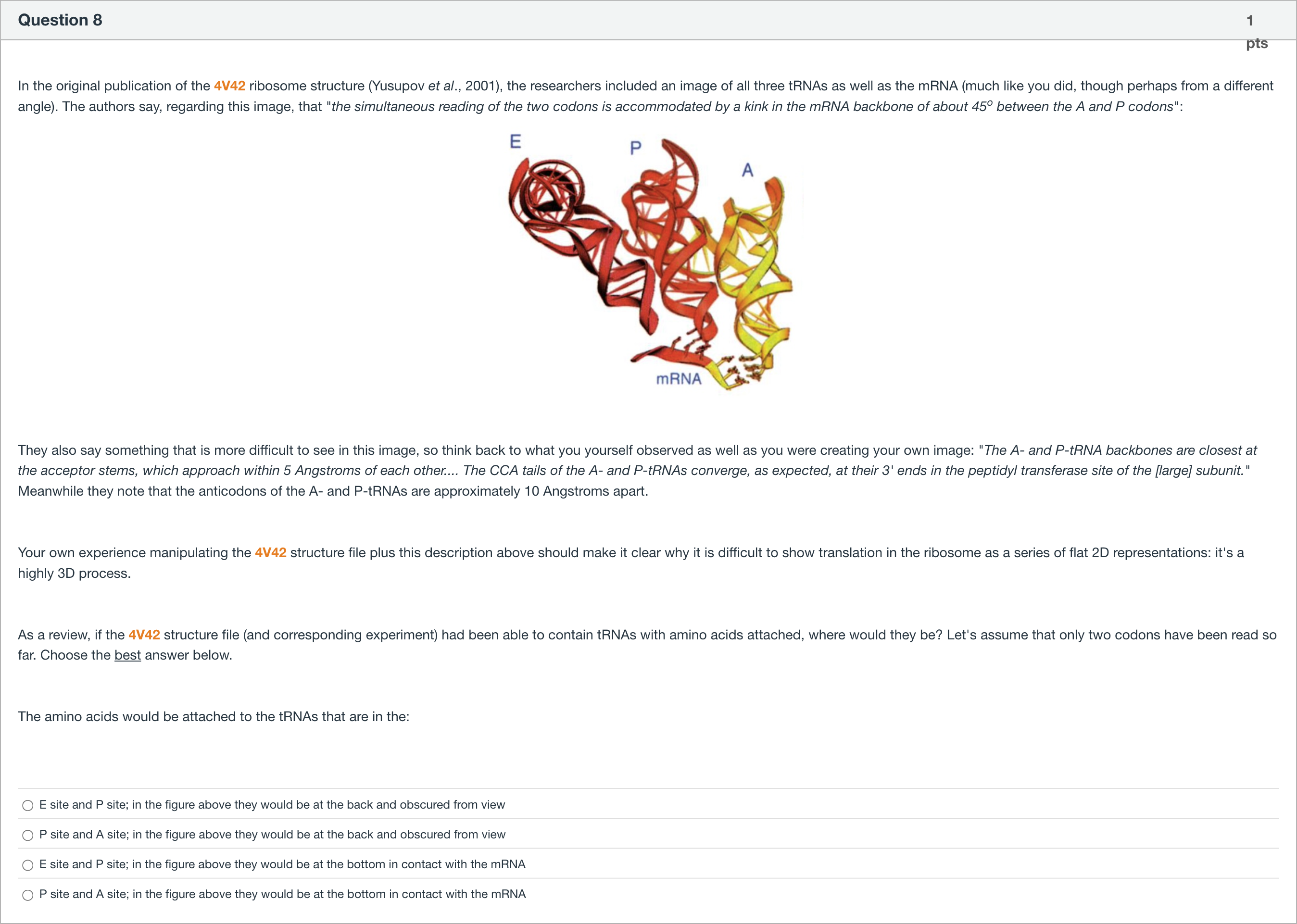
In the original publication of the 4V42 ribosome structure (Yusupov et al., 2001), the researchers included an image of all three tRNAs as well as the mRNA (much like you did, though perhaps from a different angle). The authors say, regarding this image, that "the simultaneous reading of the two codons is accommodated by a kink in the mRNA backbone of about 45o between the A and P codons": They also say something that is more difficult to see in this image, so think back to what you yourself observed as well as you were creating your own image: "The A- and P-tRNA backbones are closest at the acceptor stems, which approach within 5 Angstroms of each other.... The CCA tails of the A- and P-tRNAs converge, as expected, at their 3' ends in the peptidyl transferase site of the [large] subunit." Meanwhile they note that the anticodons of the A- and P-tRNAs are approximately 10 Angstroms apart. Your own experience manipulating the 4V42 structure file plus this description above should make it clear why it is difficult to show translation in the ribosome as a series of flat 2D representations: it's a highly 3D process. As a review, if the 4V42 structure file (and corresponding experiment) had been able to contain tRNAs with amino acids attached, where would they be? Let's assume that only two codons have been read so far. Choose the best answer below. The amino acids would be attached to the tRNAs that are in the: 单项选择题
A
E site and P site; in the figure above they would be at the back and obscured from view
B
P site and A site; in the figure above they would be at the back and obscured from view
C
E site and P site; in the figure above they would be at the bottom in contact with the mRNA
D
P site and A site; in the figure above they would be at the bottom in contact with the mRNA
登录即可查看完整答案
我们收录了全球超50000道真实原题与详细解析,现在登录,立即获得答案。
类似问题
Take this sequence and find the start site, the stop site for translation. Then write down the first 20 amino acids. (To do this scan down the DNA until you find the first ATG, then section the sequence into triplets until you find a stop codon). To translate this gene into a protein, use the above codon table. Use the 1 letter code for your answer now spaces. For example MALLS. A correct answer will have 20 uppercase letters in it
Question at position 67 Translate the template DNA sequence into protein as a ribosome would. I recommend working this out on scrap paper, then finding the answer. Template DNA: 5'-TTTAAAACACCCACCGATGTGTCATT-3'N-Met-Cys-His-CN-Lys-Phe-Cys-Gly-Trp-His-His-Ser-CN-Met-Thr-His-Arg-Trp-Val-Phe-CNo protein is madeN-Met-Thr-His-Arg-Trp-Val-Phe-Trp-C
Question at position 64 Translate the coding DNA sequence into protein as a ribosome would. I recommend working this out on scrap paper, then finding the answer. Coding DNA: 5'-GCACTATGCCCCTGCGGGGATGAGTA-3'N-Met-Pro-Leu-Arg-Gly-CN-Ala-Leu-Cys-Pro-Cys-Gly-Asp-Glu-CN-Met-Pro-Leu-Arg-Gly-Trp-CNo protein is madeN-Met-Ser-Arg-Gly-Val-Pro-Val-C
Question at position 79 In both prokaryotes and eukaryotes, how many ribosomes can translate an mRNA at a time?twothreemanyone
更多留学生实用工具
希望你的学习变得更简单
加入我们,立即解锁 海量真题 与 独家解析,让复习快人一步!