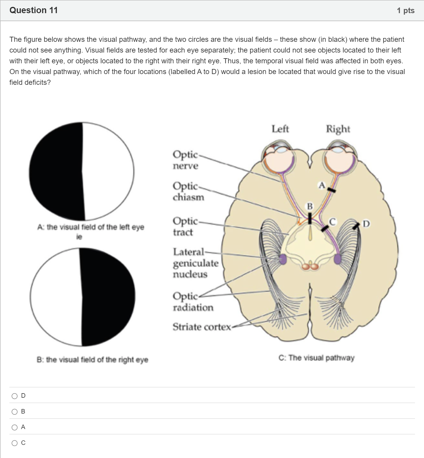
The figure below shows the visual pathway, and the two circles are the visual fields – these show (in black) where the patient could not see anything. Visual fields are tested for each eye separately; the patient could not see objects located to their left with their left eye, or objects located to the right with their right eye. Thus, the temporal visual field was affected in both eyes. On the visual pathway, which of the four locations (labelled A to D) would a lesion be located that would give rise to the visual field deficits? Single choice
A
D
B
B
C
A
D
C
Log in for full answers
We've collected over 50,000 authentic original questions and detailed explanations from around the globe. Log in now and get instant access to the answers!
Similar Questions
What is the place where the optic nerve from each each meets?
Which of the following statements is FALSE:
What is the point at which the pathway of visual information from the eye through the brain switches sides of the brain?
Question at position 48 The crossover point in the eye, where the visual information originating in the right halves of the two retinas is transmitted to the right side of the occipital lobe is called theoptic nerve.cornea.optic chiasm.fovea.
More Practical Tools for Students Powered by AI Study Helper
Making Your Study Simpler
Join us and instantly unlock extensive past papers & exclusive solutions to get a head start on your studies!